Showing posts with label Cardiac Arrest. Show all posts

Monday, May 25, 2015

Double Sequential (or Simultaneous?) Defibrillation for Refractory VF

The presence of sudden cardiac death is estimated to occur 300,000 to 350,000 annually with over 90% of such deaths as a results of ventricular fibrillation (VF). ACLS guidelines dictate that after addressing reversible causes or factors leading to the arrhythmia (hypoxia, electrolyte disturbances, mechanical factors, volume depletion), defibrillation should be performed with 360 J for monophasic defibrillators or 120-200 J for biphasic defibrillators. In a subset of patients, however, conventional means of terminating ventricular arrhythmias does not work. Energy requirements for refractory VF is controversial and, recently, the idea of double sequence defibrillation (DSD) has become a solution to refractory VF and subsequent death.

DSD is performed by attaching two sets of defibrillation pads rather than one and delivering two shocks as near simultaneously as possible, delivering electricity to the myocardial tissue in parallel pathways. The idea is that several factors affect the defibrillation threshold such as obesity, chronic lung disease, antiarrhythmic agents, decreased ejection fraction, body position/habitus, and presence of implanted internal defibrillator.

Hoch et al advocate for DSD in refractory VF. Hoch found that all five patients in the study converted to normal sinus rhythm after double sequence defibrillation at a total of 720 J. Other support for DSD come from the Cabanas paper, a retrospective case series which looked at 10 cases of refractory VF. In the paper, DSD successfully terminated 70% of refractory VF, attaining ROSC in 30% of those patients. Unfortunately, however, none of these patients survived to discharge. A contributing factor to explain the fact that there were no survivors to discharge was that DSD was performed too late. In the cases reviewed, 6.5 single shocks were given prior to DSD and in 6 of those cases, DSD was performed 35 minutes into resuscitation, which was probably too late.
           
Currently several systems around the world are using DSD for refractory VF.  Currently, we do not know the amount of joules to use for best survival. Nor do we know the correct number of pads or best pad vector. The risk/benefit profile seems very reasonable since all refractory VF leads to death. It is possible that we have finally figured out how to save these patients’ lives.

Anterior-Lateral/Anterior-Lateral

 Anterior-Lateral/Anterior-Posterior


References

  1. Chang, Mau-Song et al. Double and Triple Sequential Shocks Reduce Ventricular Defibrillation Threshold in Dogs With and Without Myocardial Infarction. Journal of the American College of Cardiology 1986; 8 (6): 1393-1405.
  2. Hoch, David H et al. Double Sequence External Shocks for Refractory Ventricular Fibrillation. JAC 1994; 23(5): 1141-1145.
  3. Zipes, Douglas P et al. Management of Patients with Ventricular Arrhythmias and the Prevention of Sudden Cardiac Death. American Heart Association, American College of Cardiology Foundation 2006.
  4. Pantridge, J. F et al. Electrical Requirements for Ventricular Defibrillation. British Medical Journal 1975; 2: 313-315.
  5. Geddes, L. A. et al. Electrical Dose of Ventricular Defibrillation of Large and Small Animals Using Precordial Electrodes. Journal of Clinical Investigation 1974; 53(1): 310-319.
  6. Adgey, A. A. J. Electrical energy requirements for ventricular defibrillation. British Heart Journal 1978; 40: 1197-1199.
  7. Cabaas, J. G. Double sequence external defibrillation in out-of-hospital refractor ventricular fibrillation: a report of ten cases. Prehospital Emergency Care 2015; 19(1): 126-130.
  8. Tacher, W. A. et al. Energy dosage for human trans-chest electrical ventricular defibrillation. New England Journal of Medicine 1974; 290: 214-215

Wednesday, March 11, 2015

Let It Flow! Intraosseous Flow Rates by Insertion Site

Intraosseous (IO) access has become increasingly popular as a ‘safety net’ for failed IV access and has become a go-to procedure in pre-hospital cases of rapidly decompensating patients. Think of the cardiac arrest patient or the hemodynamically unstable trauma patient. The IO has proven a quick and reliable way to gain access to medullary venous plexuses in long bones, which drain into systemic venous circulation. We can basically think of the bone marrow as a vein that will not collapse on us that can be accessed very rapidly, with very little training. We have infused fluids, medications, and blood products successfully through the IO. And now that increased support has grown for the use of IOs prehospitally, the question has become which site is best: Tibia (which many people seem to be most comfortable with due to prominent landmarks and distance from resuscitative efforts), humerus, or sternum. There is literature supporting, and widespread consensus for, proximal tibia as the optimal insertion site in children, but this consensus does not exist in adults. Choice often depends on comfort level of the operator and convenience of the location, but we should also consider the difference in flow rates between sites.

There are few studies comparing IO placement sites but the ones that exist compare proximal to distal tibia, tibia to humerus, and one study which compared IO infusion rates between tibia, humerus and sternum in cadavers. The first study found that IO flow rates in the proximal tibia were significantly faster with and without use of a pressure bag than flow rates the in the distal tibia. The drawback of this study was its small sample size of only 22 patients. Pasley et al did a cadaver study published 1 year ago which utilized 16 cadavers to compare flow rates and found that the sternal site had the highest and most consistent flow rate compared to the humerus and tibia. In fact the average flow rate in the sternum according to this study was 1.6x higher than the humerus and 3.1x higher than the tibia. Additionally, this study showed that the tibia had the greatest number of insertion difficulties (In 3 out of the 16 cadavers, infusion was unsuccessful after insertion and alternate tibia had to be used.) Ong et al did a study in 2009 which had very different results. This study recruited 24 patients who presented to an ED in Singapore, all patients received a tibial IO, and those who needed a second access point were given a humeral IO (which 11 patients received). This study found no significant difference between the flow rates at the tibial and humeral site in contrast to Pasley’s study which did show a significant difference between humerus and tibia with the humeral site achieving a 1.8x greater volume on average than the tibia. Small sample size is an issue in all articles existing on this subject.

It seems that there have been no conclusive studies in human or cadaver studies on best IO insertion site, but, if we believe the most recent study by Pasley et al, the sternal and humeral IO sites, in that order, have better flow rates compared to tibial placement. This higher flow rate could make a difference when rapid fluid resuscitation is imperative and could lead to better survival of our patients. A new device called the FASTResponder was released by Pyng Medical in 2013 to make the sternal IO concept easier. This device is safe on ages 12 years and older and makes site identification easy. Another benefit of the device is, unlike the IO drill system, it requires no batteries, and, anecdotally, there is less pain on fluid delivery compared to other sites. One factor we are still unclear about is if the sternal IO could pose a problem if cervical immobilization is being used in trauma patients, with chest compressions, and for some airway procedures. Pyng Medical advertises on their website that it is “safe” to use in conjunction with cervical immobilization devices and CPR. However, the drill-based EZ-IO is approved for all ages, and many providers are already comfortable with it. According to Pasley’s study, the humeral placement is second best in terms of flow rates and had less insertional difficulties. He also notes that the humeral site had the greatest variability in volumes infused from subject to subject. There doesn’t seem to be enough evidence yet to draw firm conclusions; more studies are needed with a greater number of test subjects to increase reliability.  Furthermore, outcome measures, though often difficult to study, would be nice.

References

Carness J, Russell J, Rodrigo M, et al. Fluid Resuscitation Using the Intraosseous Route: Infusion with Lactated Ringer’s and Hetastarch. Military Medicine 2012; 2:222.

Ong M, Chan Y, Jen J, Ngo A. An observation prospective study comparing tibial and humeral intraosseous access using the EZ-IO. Amer Journal of Emergency Medicine 2009; 27, 8-15. 

Pasley J, Miller C, Dubose J, et al. Intraosseous Infusion Rates under High Pressure: A Cadaveric Comparison of Anatomic Sites. Distribution A: Approved for Public Release 2014: Case Number 88ABW-2014-1139.

Tan B, Chong S, Koh Z, Ong M. EZ-IO in the ED: an observational, prospective study comparing flow rates with proximal and distal tibia intraosseous access in adults. Amer Journal of EM 2012;30(8):1602-6.

Friday, January 23, 2015

More Than Just Shock Value?

*Note - this discussion is only pertinent to modern biphasic defibrillators with self adhesive electrodes applied only anterior/posterior or anterior/lateral, with the compression provider wearing two pairs of gloves (double-gloving), with a maximum defibrillation energy of 360J.*
We have all heard the chant, “I’m clear, you’re clear, we’re all clear,” prior to a provider double, triple, sometimes quadruple checking him or herself before pushing that magic red button with the white lightning bolt - “shock”!  Recent literature has spurred quite the discussion on hands-on defibrillation (HOD) - CPR where compressions continue throughout the defibrillation - as it is widely known that interruptions in chest compressions lead to poor patient outcomes and are all too common, for example, during intubation, providing ventilations, AED analyzing, charging, and during defibrillation shocks.  This pearl is meant to provide a very brief explanation of what your risks might be, what protection devices you might use, anecdotal and published accounts on HOD, and suggestions for your clinical practice.
There are numerous factors in regards to energy and the effect it may have on the provider during HOD.  Energy is the product of voltage, current and time.  Neither factors, independently, are sufficient in inducing damaging effects.  For example, several thousand volts are experienced during static electricity, although the current is very low.  Current is determined by the resistance between the electrodes of the defibrillator, the electrode gel, the gel-skin contact, and the tissue resistance.  Glove integrity, skin moisture and the actual current pathway will determine the amount of escape current.  Biphasic defibrillators provide voltages up to approximately 2200 V over approximately 15-20 msec.  The maximum permissible leakage current, per the International Commission on Non-Ionizing Radiation Protection is 1mA; the threshold for perception is 2.5-4.0 mA; and pain is experienced at 6-10 mA.2  Sullivan and Chapman studied the voltage-current curves for gloves.  They note the international safety standard on 1mA and explain that at this level, it would take 1-3 seconds of current flow to induce VF in <5% of the population.  While defibrillation shocks are usually less than 20 msec, even if the pulse is timed appropriately in the rescuer’s cardiac cycle, as much as 500 mA would be required to induce VF.1 For reference, the current exposure from a home body fat monitoring scale is 500 uA.
In one of the most exciting studies, Lloyd et al measured current between “rescuers” and patients undergoing cardioversion at up to 360 J and found the highest current leak measured was 907 uA,4 with no “rescuers” experiencing a “shock.”  Neumann et al found HOD was safely performed on pigs by rescuers, HOD shortened pauses during CPR, and it more quickly restored coronary perfusion pressure.4  Kurz and Sawyer, in their letter to the editor of Resuscitation, advocate eliminating effects of no-flow time, perhaps by using HOD.7 Dr. Scott Weingart writes that in the 4 years that he and his colleagues have been performing HOD, there have been no rescuer complications, although occasional perceptions of tingling have been reported.  He himself reported arm soreness after 3 shocks, all at 360 J with the electrode pads notably in the anterior/anterior position.
In opposition, Lemkin et al derive an equation called the rescuer-received dose, to try to better qualify defibrillation risk.  Noting that energy values greater than 1 J reportedly can cause VF, they deem HOD unsafe as values above 1 J were calculated in their cadaver study, though effects of gloves were not accounted for.  Two studies from the UK found that medical examination gloves do not provide rescuer safety and even demonstrate further glove breakdown of the gloves worn by rescuers who perform compressions.  According to Sullivan and Chapman, HOD with medical examination gloves will produce no sensation at all unless the gloves completely break down.1  
Although there are no reported fatalities or serious consequences to rescuers performing HOD under ideal conditions - using a biphasic defibrillator with electrodes placed appropriately, with rescuers double gloved - we should take note that any change to a safety protocol should not be undertaken without ensuring rescuers' safety.  I have personally performed HOD, as have my colleagues in the emergency department.  While none of us have experienced any detrimental consequences or even the reported tingling, considering the literature, perhaps we should currently hold off on changing our protocols to mandate hands-on defibrillation.  Protocols that need to be changed or followed are as follows:

  • High quality CPR remains of utmost importance.  Set a metronome at 100 beats per minute and compress the chest to 1.8” (or as close to it as possible) every time.
  • Have no interruptions in chest compressions - not for intubation, not for starting an IV, not for inserting a central line, not for transporting, and not for charging the defibrillator!
                  
The use of HOD needs to reflect your clinical decision made in the best interest of you, your co-rescuers, and your patient.  If you chose to do so, please double-glove, please place the electrodes anterior/posterior, and communicate your practice to your colleagues.    

References

1.  Sullivan JL, Chapman FW. Will medical examination gloves protect rescuers from defibrillation voltages during hands-on defibrillation? Resuscitation. 2012 Dec;83(12):1467-72. doi: 10.1016/j.resuscitation 2012.07.031. Epub 2012 Aug 25. PubMed PMID: 22925991

2.  Petley GW, Cotton AM, Deakin CD. Hands-on defibrillation: theoretical and practical aspects of patient and rescuer safety. Resuscitation. 2012 May;83(5):551-6. doi: 10.1016/j.resuscitation.2011.11.005. Epub 2011 Nov 15. Review. PubMed PMID: 22094984.

3.  Sullivan JL. Letter by Sullivan regarding article, "Hands-on defibrillation: an analysis of electrical current flow through rescuers in direct contact with patients during biphasic external defibrillation". Circulation. 2008 Dec 2;118(23):e712; author reply e713. doi: 10.1161/CIRCULATION AHA.108.803718. PubMed
PMID: 19047587.

4.  Lloyd MS, Heeke B, Walter PF, Langberg JJ. Hands-on defibrillation: an analysis of electrical current flow through rescuers in direct contact with patients during biphasic external defibrillation. Circulation. 2008 May 13;117(19):2510-4. doi: 10.1161/CIRCULATION AHA.107.763011. Epub 2008 May 5. PubMed PMID: 18458166.

5.  A note of caution on the performance of hands-on biphasic defibrillation. Weingart SD. Resuscitation. 2013 Mar;84(3):e53. doi: 10.1016/j.resuscitation.2012.12.014. Epub 2012 Dec 22. PMID: 23266533

6. Lemkin DL, Witting MD, Allison MG, Farzad A, Bond MC, Lemkin MA. Electrical exposure risk associated with hands-on defibrillation. Resuscitation. 2014 Oct;85(10):1330-6. doi: 10.1016/j.resuscitation.2014.06.023. Epub 2014 Jun 30. PubMed PMID: 24992873.

7.  Petley GW, Deakin CD. Do clinical examination gloves provide adequate electrical insulation for safe hands-on defibrillation? II: Material integrity following exposure to defibrillation waveforms. Resuscitation. 2013 Jul;84(7):900-3. doi: 10.1016/j.resuscitation.2013.03.012. Epub 2013 Mar 16. PubMed PMID: 23507465.

8.  Deakin CD, Lee-Shrewsbury V, Hogg K, Petley GW. Do clinical examination gloves provide adequate electrical insulation for safe hands-on defibrillation? I: Resistive properties of nitrile gloves. Resuscitation. 2013 Jul;84(7):895-9. doi: 10.1016/j.resuscitation.2013.03.011. Epub 2013 Mar 16. PubMed PMID: 23507464.

Saturday, December 27, 2014

End-Tidal Carbon Dioxide Monitoring in the Resuscitation of Critically Ill & Injured

Case:
73 yo male with PMH of CAD s/p CABG, hypertension, other medical history unavailable. C/C sudden cardiac witnessed by his wife at approximately 10:55 am. CPR instructions given to his wife over the phone by the 911 operator. Police arrived minutes later to continue CPR with an AED. The BLS team arrived shortly after and assisted with the resuscitation using an AED, BVM, OPA and supplemental oxygen. ALS arrived 14 minutes after the 911; BLS reported 3 AED defibrillations prior to ALS arrival.

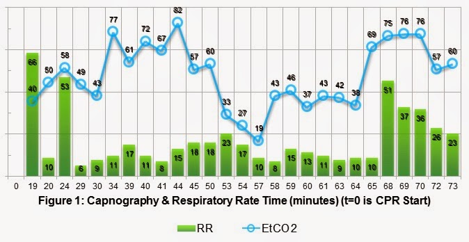
Initial ECG is coarse ventricular fibrillation and the patient was defibrillated at 360J by ALS. High-Quality CPR continued. Vascular access established with a right proximal humoral IO, 1mg of epinephrine given and repeated every 4 minutes during CPR. Paramedics intubated the patient without an interruption in chest compressions; initial EtCO2 is 40 mmHg. 2 minutes later, the patient is noted to be in refractory VFIB despite serial defibrillations and anti-dysrhythmics. A total of 14 defibrillations, amiodarone, magnesium and lidocaine were required to convert the VFIB to an organized pulseless sinus ECG rhythm with a QRS width of 160 msec. The patient also received calcium and sodium bicarbonate. Return of spontaneous circulation (ROSC) was noted 38 minutes into the resuscitation. This patient received approximately 1200 cc of crystalloid IVF, atropine and push dose pressors (1:100k Epi) to maintain hemodynamics during transfer to the ED. Pulses were lost during transport for 8 minutes and required CPR and additional epinephrine. Below is a plot of his EtCO2 and respiratory rate versus time. ROSC#1 @ t=38 minutes and ROSC#2 at t=65 minutes.


Kodali and colleagues recently published an excellent review of the usefulness of capnography in Care of the Critically Ill and Injured. The literature search was quite extensive looking at peer review papers from 1960 to 2014, covering primary research, case reports and other review papers. Figure 2 is a summary of the different clinical applications. Conformation of endotracheal intubation is quoted in Kodali's paper as both 100% sensitive and specific, with 3 decades of data for detecting correct tube placement. This has been known for a while in EM and nice to repeat as often as possible because few tests have that level of certainty. Waveform Capnography is the most definitive evidence of correct endotracheal tube placement, thus eradicating the unrecognized esophageal intubation.


Capnography has been demonstrated to reflect the patients cardiac output (CO) during a resuscitation based on the height of the waveform. The greater the CO the more CO2 is off loaded in the lungs and measured on exhalation. Current Evidence  demonstrate EtCO2 levels less than 10 mmHg during chest compression is not likely to generate ROSC, so every effort should be made to maximize the quality of CPR and treat reversible causes of arrest.

A certain level of prognostication or prediction is also gained by the routine use of capnography during CPR. Abrupt increases in EtCO2,  generally a jump greater than 10-20 mmHg, is a marker of ROSC, and, conversely, refractory EtCO2 values less than 10 mmHg has identified 100% of patients who were unsuccessfully resuscitated. Kodali found that the cumulative max EtCO2 > 20 mmHg at all time points measured between 5 and 10 minutes post-intubation best predicted ROSC (sensitivity of 88%, specificity 77%). EtCO2 is a valuable tool in real-time decision-making during resuscitation.

Other uses of capnography include monitoring of airway patency and respiratory rates. The waveform capnograph and the ability to set warning alarms will instantly alert providers to apneic conditions, such as obstruction or displacement. In clinical situations where a patient is sedated or obtunded, EtCO2 will herald hypoventilation or apnea much sooner than traditional SpO2 monitoring.

Back to our case, after reviewing figure #1, we can apply all the previously mentioned key points to capnography. EtCO2 definitively confirmed ETT placement. ROSC was predicted at t=10 minutes by an EtCO2 value greater than 20 mmHg. High-Quality CPR was performed while reversible causes of the arrest were managed. ETT patency was maintained throughout the encounter and transfer to the ED. This would have been a great case to use 720 J Double Sequential Defibrillation (DSD) for refractory ventricular fibrillation.

References:

1.     Goto, Y; etal. Termination-of-resuscitation rule for emergency department physicians treating out-of-hospital cardiac arrest patients: an observational cohort study. Critical Care. 2013;17:R235.
2.     Kodali, BS; etal. Capnography during cardiopulmonary resuscitation: Current evidence and future directions. J Emerg Trauma Shock. 2014;7(4):332-340.
3.     Meaney, PA; etal. Cardiopulmonary Resuscitation Quality: Improving Cardiac Resuscitation Outcomes Both Inside and Outside the Hospital. A Consensus Statement From the American Heart Association Endorsed by the American College of Emergency Physicians and the Society of Critical Care Medicine. Circulation. 2013;128:417-435.
4.     Neumar, RW; etal. Part 8: Adult Advanced Cardiovascular Life Support: 2010 American Heart Association Guidelines for Cardiopulmonary Resuscitation and Emergency Cardiovascular Care. Circulation. 2010;122[suppl 3]:S729S767.