Sunday, December 28, 2014

2015: The Death of Longboards (Hopefully)

Myths in medicine take too long to go away.  Longboards are yet another modality that serve no purpose except to harm our patients.  Luckily this unnecessary tool used by EMS is going away around the world.  Many states and cities have completely stopped using longboards for ALL patients.  These places include areas within Connecticut, Los Angeles, Kansas, Oregon, Missouri, Houston, New Mexico,.etc.  No matter what your injuries are, in many regions throughout the world, you will not be placed on a longboard, because they are not being used at all.

These devices have hurt our patients since they offer no benefit, yet we continue to use them in our region.  The misconceptions about these devices are enormous, yet the science tells us the following...

Longboards:
1.      Worsen the pain of patients resulting in more unnecessary imaging tests and more radiation exposure.
2.      Cause respiratory compromise/decreased pulmonary function by lying patients flat.
3.      Delay on-scene time for trauma patients.
4.      Result in pressure sores for patients by rapid tissue breakdown from the board.
5.      Increase the risk of aspiration.

Unfortunately, we continue to have folks who spread misconceptions about these devices, which prevent us from moving forward with evidence based medicine.  Luckily, a lot of places are ignoring these folks and moving forward.  Some of the incorrect EMS statement that we have heard are:
1.      The DOT makes me put everyone on a longboard.
2.      I will get my license/certification taken away if I don’t use a longboard.
3.      The DHSS does not allow patients to be brought to hospitals without a longboard.
4.      If someone else puts a patient on a longboard, I cannot take the patient off.
5.      It splints the back.  (No, in fact it was only designed to help extricate patients.)
6.      I will get sued if I don’t put someone on a longboard

These are all ridiculous, and it is great that many places around the country are moving forward with the science.  Lets make 2015 the year we get rid of these terrible devices in New Jersey and around the country.

Following the science, in January 2015, we will be telling EMS providers that they do not need to place anyone on a longboard that is brought into our hospital.  Please join us in getting rid of this outdated modality and provide the same information to EMS.

References:

1. Chan D, Goldberg R, Tascne A, et al. The effect of spinal immobilization on healthy volunteers. Ann Emerg Med. 1994;23:48-51.
2. March JA, Augband SC, Brown LH. Changes In Physical Examination Caused By Use Of Spinal Immobilation. Prehospital Emerg Care. 2002; 6: 421-424.
3. Schriger DL, Larmon B, LeGarrick T, et al. Spinal immobilization on a flat backboard: Does it result in neutral position of the cervical spine? Am J Emerg Med. 1991;20:878-81.
4. Schafermeyer RW, Ribbeck BM, Gaskins J, et al. Respiratory effects of spinal immobilization in children. Ann Emerg Med. 1991;20:1017-1019.
5. Bauer D, Kowalski R. Effect of spinal immobilization devices on pulmonary function in the healthy nonsmoking man. Ann Emerg Med.-1988; 17:915-8.
6. Barney RN, Cordell WH, Miller E. Pain associated with immobilization on rigid spine boards (Abstract). Ann Emerg Med.1989; 18:918.
 7. Chan D, Goldberg, RM,  Jennifer Mason, J et al., Backboard Versus Mattress Splint:  A Comparison Of Symptoms. The Journal of Emergency Medicine. 1996. 14:193-298.
8. Totten VY, Sugarman DB, Respiratory Effects Of Spinal Immobilization. Prehosp Emerg Care 1999;3:347-352
9.  Hauswald M,  McNally T. Confusing Extrication with Immobilization: The Inappropriate Use of Hard Spine Boards for Interhospital Transfers. Air Med J. 2000; 19: 126-127
10. Hauswald M,Braude D.Spinal immobilization in trauma patients: is it really necessary?_Current Opinion in Critical Care 2002;8:566–70.
11. Hauswald M,Ong G,Tandberg D,Omar Z. Out-of-hospital spinal immobilization:  its effect on neurologic injury.  Academic Emergency Medicine 1998;5:214-219.
12. S. Abram S, Bulstrode C. Routine spinal immobilization in trauma patients: What are the advantages and disadvantages? The Surgeon. 2010;8:218–222.
13. Connell RA, Graham CA, Munro PT. Is spinal immobilization necessary for all patients sustaining isolated penetrating trauma? Injury. 2003;34: 912–914.
14. Kaups KL, Davis JW. Patients With Gunshot Wounds To The Head Do Not Require Cervical Spine Immobilization And Evaluation. J Trauma. 1998; 44:865– 867.
15. Haut ER,  Efron DT,  Adil H, Haider AH et al. Spine Immobilization in Penetrating Trauma: More Harm Than Good? The Journal of Trauma. 2010;  68.
16. Cornwell EE, Chang DC, Bonar JP, et al. Thoracolumbar immobilization for trauma patients with torso gunshot wounds: is it necessary? Arch Surg. 2001;136:324 –327.
17. Hauswald M, Ong G, Tandberg D, Omar Z. Out-of-hospital spinal immobilization: its effect on neurologic injury. Acad Emerg Med. 1998;5:214 –219.
18. Kaups KL, Davis JW. Patients with gunshot wounds to the head do not require cervical spine immobilization and evaluation. J Trauma. 1998;44:865–867.
19. Mark Hauswald, MD, Darren Braude, MD, MPH .Diffusion of Medical Progress: Early Spinal Immobilization in the Emergency Department. Academic Emergency Medicine 2007; 14:1087–1089.
Saturday, December 27, 2014

Journal Club - December 29, 2014


  • What:  Weekly Journal Club
  • Where:  MONOC Education Building, 1415 Wyckoff Road, Ground Floor, Wall Township, NJ
  • When:  Monday, December 29, 2014 at 10:00 am
  • Tranexamic acid for traumatic brain injury: a systematic review and meta-analysis.  http://www.ncbi.nlm.nih.gov/pubmed/25447601.  Proof that we should get rid of the relative contraindication for isolated head trauma?
  • Use of High-Flow Nasal Cannula Oxygen Therapy to Prevent Desaturation During Tracheal Intubation of Intensive Care Patients with Mild-to-Moderate Hypoxemia.  http://www.ncbi.nlm.nih.gov/pubmed/25479117.  If you're not putting a high-flow nasal cannula on every critically ill patient, you're doing something wrong!  NO DESAT.
Live-tweeting of the journal club @CCareAnywhere.

End-Tidal Carbon Dioxide Monitoring in the Resuscitation of Critically Ill & Injured

Case:
73 yo male with PMH of CAD s/p CABG, hypertension, other medical history unavailable. C/C sudden cardiac witnessed by his wife at approximately 10:55 am. CPR instructions given to his wife over the phone by the 911 operator. Police arrived minutes later to continue CPR with an AED. The BLS team arrived shortly after and assisted with the resuscitation using an AED, BVM, OPA and supplemental oxygen. ALS arrived 14 minutes after the 911; BLS reported 3 AED defibrillations prior to ALS arrival.

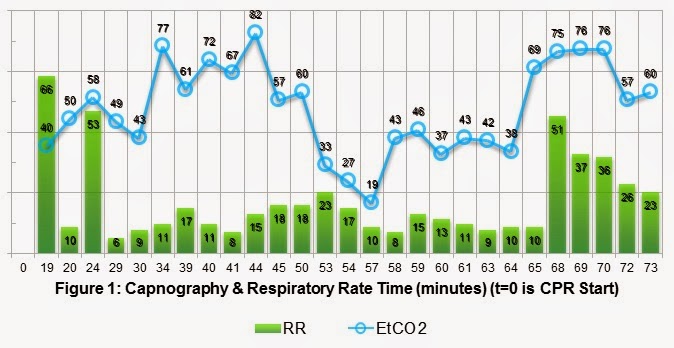
Initial ECG is coarse ventricular fibrillation and the patient was defibrillated at 360J by ALS. High-Quality CPR continued. Vascular access established with a right proximal humoral IO, 1mg of epinephrine given and repeated every 4 minutes during CPR. Paramedics intubated the patient without an interruption in chest compressions; initial EtCO2 is 40 mmHg. 2 minutes later, the patient is noted to be in refractory VFIB despite serial defibrillations and anti-dysrhythmics. A total of 14 defibrillations, amiodarone, magnesium and lidocaine were required to convert the VFIB to an organized pulseless sinus ECG rhythm with a QRS width of 160 msec. The patient also received calcium and sodium bicarbonate. Return of spontaneous circulation (ROSC) was noted 38 minutes into the resuscitation. This patient received approximately 1200 cc of crystalloid IVF, atropine and push dose pressors (1:100k Epi) to maintain hemodynamics during transfer to the ED. Pulses were lost during transport for 8 minutes and required CPR and additional epinephrine. Below is a plot of his EtCO2 and respiratory rate versus time. ROSC#1 @ t=38 minutes and ROSC#2 at t=65 minutes.


Kodali and colleagues recently published an excellent review of the usefulness of capnography in Care of the Critically Ill and Injured. The literature search was quite extensive looking at peer review papers from 1960 to 2014, covering primary research, case reports and other review papers. Figure 2 is a summary of the different clinical applications. Conformation of endotracheal intubation is quoted in Kodali's paper as both 100% sensitive and specific, with 3 decades of data for detecting correct tube placement. This has been known for a while in EM and nice to repeat as often as possible because few tests have that level of certainty. Waveform Capnography is the most definitive evidence of correct endotracheal tube placement, thus eradicating the unrecognized esophageal intubation.


Capnography has been demonstrated to reflect the patients cardiac output (CO) during a resuscitation based on the height of the waveform. The greater the CO the more CO2 is off loaded in the lungs and measured on exhalation. Current Evidence  demonstrate EtCO2 levels less than 10 mmHg during chest compression is not likely to generate ROSC, so every effort should be made to maximize the quality of CPR and treat reversible causes of arrest.

A certain level of prognostication or prediction is also gained by the routine use of capnography during CPR. Abrupt increases in EtCO2,  generally a jump greater than 10-20 mmHg, is a marker of ROSC, and, conversely, refractory EtCO2 values less than 10 mmHg has identified 100% of patients who were unsuccessfully resuscitated. Kodali found that the cumulative max EtCO2 > 20 mmHg at all time points measured between 5 and 10 minutes post-intubation best predicted ROSC (sensitivity of 88%, specificity 77%). EtCO2 is a valuable tool in real-time decision-making during resuscitation.

Other uses of capnography include monitoring of airway patency and respiratory rates. The waveform capnograph and the ability to set warning alarms will instantly alert providers to apneic conditions, such as obstruction or displacement. In clinical situations where a patient is sedated or obtunded, EtCO2 will herald hypoventilation or apnea much sooner than traditional SpO2 monitoring.

Back to our case, after reviewing figure #1, we can apply all the previously mentioned key points to capnography. EtCO2 definitively confirmed ETT placement. ROSC was predicted at t=10 minutes by an EtCO2 value greater than 20 mmHg. High-Quality CPR was performed while reversible causes of the arrest were managed. ETT patency was maintained throughout the encounter and transfer to the ED. This would have been a great case to use 720 J Double Sequential Defibrillation (DSD) for refractory ventricular fibrillation.

References:

1.     Goto, Y; etal. Termination-of-resuscitation rule for emergency department physicians treating out-of-hospital cardiac arrest patients: an observational cohort study. Critical Care. 2013;17:R235.
2.     Kodali, BS; etal. Capnography during cardiopulmonary resuscitation: Current evidence and future directions. J Emerg Trauma Shock. 2014;7(4):332-340.
3.     Meaney, PA; etal. Cardiopulmonary Resuscitation Quality: Improving Cardiac Resuscitation Outcomes Both Inside and Outside the Hospital. A Consensus Statement From the American Heart Association Endorsed by the American College of Emergency Physicians and the Society of Critical Care Medicine. Circulation. 2013;128:417-435.
4.     Neumar, RW; etal. Part 8: Adult Advanced Cardiovascular Life Support: 2010 American Heart Association Guidelines for Cardiopulmonary Resuscitation and Emergency Cardiovascular Care. Circulation. 2010;122[suppl 3]:S729S767.

Tuesday, December 16, 2014

Hydrofluoric Acid Burns

A 32 year old gentleman was welding stainless steel and sustained a 75% concentration hydrofluoric acid burn to the right hand.  Upon initial assessment, vital signs are unremarkable with a RR-12 @ 99% RA and BP 128/86.  On physical exam there are no visible signs of damage to the right hand and he seems to be neurovascularly intact.  Upon auscultation of the heart and lungs, diffuse expiratory wheezing is noted.  A 12 lead EKG is obtained and pictured below.  What are your initial management options and continuing concerns throughout treatment of this patient?


Hydrogen fluoride and its aqueous form, hydrofluoric acid, are utilized in many manufacturing processes.  Uses include: cleaning products, oil refining, Teflon production, aluminum production, and etching of carbon and stainless steels as well as ceramics.  Exposures most commonly seen involve explosions of Teflon containing compounds, deployment of automated fire suppression systems, and exposure to cleaning solutions.
The high lipid solubility of HF allows it to penetrate tissues rapidly.  Immediate exposure may result in a painless burn, as the substance interferes with nervous system function.  This could delay contact of emergency services and also mask the severity of exposures. Extreme pain ensues as the chemical begins to interact with ions in the tissues.  The fluoride ion in the compound has a high electronegativity.  This quality imparts a high affinity for positively charged ions in the body, allowing HF to readily interact with calcium and magnesium in the body.  The combination of hypocalcemia and hypomagnesemia promotes prolongation of the QT interval, ventricular arrhythmias, tetany, and seizures.  HF burns involving greater than 2% body surface area or HF with a concentration of greater than 20% have the highest likelihood of causing systemic toxicity and fatal arrhythmia.
As with most chemical exposures, copious irrigation is encouraged.  In addition to irrigation, literature has shown improved immediate pain relief and long-term outcomes with application of topical 2.5% calcium gluconate.  If calcium gel is not readily available, it can be made by mixing 3.5 g of calcium gluconate powder with approximately 5 ounces of water soluble surgical lubricant.  For continued pain after topical application, 5% calcium gluconate subcutaneous injections may be utilized for local infiltration.   The appropriate dose is 0.5 mL per cm² burn area.  Local infiltration is not recommended for digits.  Up to 40 mL of 10% calcium gluconate can be given intra-arterially or intravenously with an inflated blood pressure cuff to localize the treatment if infiltration is not effective or not an option.
In the case presented, cardiac monitoring and topical calcium application are the most important management factors in the pre-hospital setting.  A seemingly mild case of an HF burn to the hand could decompensate quickly due to cardiac dysrhythmia.  Keep in mind that 2% BSA can include a burn only involving the hand!  In a hospital setting, continuous cardiac and electrolyte monitoring guide management.  Calcium can be administered intravenously in a 1000 mg dose infused over 2 minutes.  Magnesium replacement of 4g IV given over 20 minutes is also recommended. 

References
1.         Nathanson L A, McClennen S, Safran C, Goldberger AL. ECG Wave-Maven: Self-Assessment Program for Students and Clinicians. http://ecg.bidmc.harvard.edu.  Accessed 16 December 2014.
2.         Höjer J, Personne M, Hultén P, Ludwigs U. Topical treatments for hydrofluoric acid burns: a blind controlled experimental study. J Toxicol Clin Toxicol. 2002;40(7):861-866. http://www.ncbi.nlm.nih.gov/pubmed/12507055.
3.         Lewis N, Lewand M, Howland N, Hoffman R, Goldfrank L, Flommenbaum L; Goldfrank’s Toxicologic Emergencies, Ninth Edition; 9th ed; 2010. 787-803.
4.         Wu M-L, Yang C-C, Ger J, Tsai W-J, Deng J-F. Acute hydrofluoric acid exposure reported to Taiwan Poison Control Center, 1991-2010. Hum Exp Toxicol. 2014;33(5):449-454. doi:10.1177/0960327113499165.

Tuesday, December 9, 2014

Journal Club - December 15, 2014


  • What:  Weekly Journal Club
  • Where:  Newark Beth Israel Medical Center, D-11, 201 Lyons Avenue, Newark, NJ
  • When:  December 15, 2014 at 2:00 pm (note the later time!)
We have an exciting lineup of articles this week!  Come join us to discuss them.

Live-tweeting of the journal club @CCareAnywhere.

Ebola - Just the Facts!

Editor note:  Thankfully, a lot of the hype has already passed on this disease.  This is the clinical pearl written a month ago, prior to the blog.  Unfortunately, I don't think we've heard the last about Ebola, as it still comes up.

Before I start this Clinical Pearl, let me say that Ebola is a scary disease, but, with any disease, we must put science and fact above panic and rumor.  People ask… am I worried about Ebola for the general population?  The answer is no… I am worried about diseases that are likely to kill the general population, such as heart disease, stroke, cancer, trauma, distracted driving, COPD, etc..  If we were to put Ebola into perspective and spend the amount of time discussing it based on the likelihood it will kill any of us compared to the previous diseases…  Well, we would never bring it up.  Unfortunately, too much confusion and too many myths surround this filovirus.  People often say, “Ebola is a lot more likely to kill someone than the diseases I just mentioned.”  That is completely untrue.  In fact, if you took patients with STEMIs or cancer and put them in areas of Ebola outbreaks, which are remote areas of Liberia, Sierra Leone, Nigeria, Senegal and Guinea, the mortality would probably be higher than Ebola is now.  Any disease process in remote areas yields a high mortality rate because of unavailable medical resources.  The actual mortality rate of Ebola in West Africa based on the first 4507 recent cases is 70 percent.  The mortality rate in hospitalized patients is 64.7 percent and 56 percent in health care workers.  So, even without substantial medical care (intravenous fluids), 30 percent of all patients survive and 44 percent of healthcare workers survive.  What is the mortality rate of Ebola patients who contracted it in the United States?  ZERO percent.  Although we have given plasma transfusions and monoclonal antibodies to these patients, we have no evidence that we help any patient beyond supportive care of IV fluids.  Even the one patient who was transferred to Germany, in septic shock with significant hypotension, received 30 liters of fluids and survived.  In the first 9 months of the recent outbreak, we know a couple of things.  The most common presenting symptoms are like anything else:  Fever (87%), fatigue (76%), decreased appetite (65%), vomiting (67%), and diarrhea (65%).  Less than five percent of patients had unexplained bleeding.  So, fever is not present in 13 percent of patients, which makes it the most common yet an unreliable finding to screen patients.  These symptoms could explain many diseases which are much more likely than Ebola.
            The transmissibility of diseases is explained by the R0  factor.  From the first 4507 patients during the recent outbreak with Ebola or probable Ebola, this factor is 1.7-2.  The Sierra Leone type is the highest.  To put this into perspective, measles is 17-19. This represents the number of patients who will contract the disease from one individual without isolation.  The incubation period is 11.4 days and the rate of conversion to a positive Ebola test is within 4 days.  Of the twenty-seven outbreaks since 1967, none have resulted in a pandemic.
            So if mortality is not as bad as we initially thought, many people are afraid of Ebola because they never thought the disease would come to the United States.  This is not the first time we have seen Ebola in the United States.  In fact it was predicted that this was going to happen.  The book “The Hot Zone” by Richard Preston in 1992 predicted the reemergence of Ebola based on a monkey outbreak in Reston, Virginia (about 10 miles from Washington, D.C.).  On Oct 2, 1989, 100 monkeys were shipped via Amsterdam through Tokyo, Tipai to New York City.  They traveled down I-95 to Reston, Virgina.  On November 1, 1989, the monkeys began dying and were incorrectly diagnosed with Simian Hemorrhagic Fever, which turned out to be Ebola Virus.  The Level 4 Biosafety Lab at Fort Dietrick correctly analyzed the tissues of the monkey.  Interestingly, although no protection was initially used in Reston, not one person contracted Ebola from the sick monkeys.  The monkeys were euthanized and significant disinfection was performed over the next 11 days.
            Another fear is the amount of personal protective equipment (PPE) one must wear around these patients.  In fact, this is really no different than any other potentially infectious disease when patients are acutely ill with diarrhea or vomiting.  In many ways, it is less since Ebola is not naturally aerosolized.  I would not come into contact with bodily fluid of any acutely ill person without universal precautions. Ebola is no different in terms of many diseases except it is less transmissible than many.  We should be observing the same universal precautions in all patients.
            Quarantining patients has been a source of great debate.  We have marked 21 days in patients as a “magical number.”  Yet this is not true.  Five percent of patients may show symptoms past the 21 day mark.  However we must follow science and folks who have dealt with this process for a long time.  Doctors without Borders is one of the amazing agencies that have dealt with this disease for years and based recommendations on science on fact.  Quarantining is not necessary unless patients are symptomatic.  Of note they also agree that, “Ebola is a hard disease to catch.”  Unless you are in contact with diarrhea or feces or vomiting, no real risk exists.  I would add that you should not come into contact with these bodily fluids as a healthcare worker and simple gloves are not enough.  I have heard of folks going into rooms with a mask and gloves, but, like all patients, this is not universal precaution, and the mask does nothing for Ebola, which is not aerosolized.
            The host of Ebola seems to be the fruit bat, and coming into contact with patients who have the highest viral load or people who have recently died poses the greatest risk of disease transmission.  Additionally, other species which are taking secretions of tissues from fruit bats seem to be at risk as well, such as apes.
            Finally, my advice is to keep this virus in perspective.  Worry about diseases which result in bad outcomes every day.  Continue to use the same precautions that hopefully you have been using for years and do not believe rumors.  Use science and the community of infectious disease folks and evidence based articles to guide you.

Ebola Virus Disease in West Africa — The First 9 Months
of the Epidemic and Forward Projections NEJM Oct 16, 2014

Transmission dynamics and control of Ebola virus disease (EVD): A Review

BMC Medicine 2014, 12:196 Oct 2014

Monday, December 1, 2014

Intubation in Head Trauma

You arrive on scene of a 17 year old male pedestrian struck, unknown medical history.  A friend who was with the patient reports that they were returning from getting dinner when he was struck by a vehicle while riding his bicycle.  The friend denies any alcohol or other intoxicating substances.  The patient is combative, fighting with rescuers, and pulling at his cervical collar.  He has evidence of head trauma externally with scalp bleeding controlled with direct pressure.  You suspect that this patient has a traumatic brain injury and needs airway management to assist in his care.  You administer 4 mg midazolam intranasal to obtain mild sedation to assist with patient care.  Approximately two minutes later, the patient is more amenable to patient care efforts.  You establish IV access, place the patient on high-flow nasal cannula, and call medical control for rapid sequence intubation orders for this approximately 70 kg patient.  The physician orders 150 mcg fentanyl IVP, 100 mg ketamine IVP, and 100 mg succinylcholine IVP.  What about lidocaine?  What about defasciculating doses of a non-depolarizing paralytic?  What about the contraindication for ketamine in head trauma?

For decades, physicians and prehospital providers have been taught many myths about intubation and intracranial pressures:  Give lidocaine prior to intubation, don’t give succinylcholine without a defasciculating dose, and never give ketamine for a head injury. 

What about pretreatment for the adrenergic response to intubation?  Clinical pearl #25 addresses the use of lidocaine as pretreatment in trauma airways.  The bottom line is that some studies show that it blunts the adrenergic response to intubation and others show no significant difference.  In fact, many of the studies were actually focused on deep tracheal suctioning of already-intubated patients.  For this reason, it is not routinely recommended that lidocaine be given prior to RSI in head injury. 

If attempting to decrease intracranial pressure (ICP) and the adrenergic response to intubation (including increased HR and BP), the better medications to use are opioids, such as fentanyl 2-3 mcg/kg, or esmolol 2 mg/kg, a short-acting beta-blocker.  Fentanyl was superior to lidocaine or placebo in blunting the increase in blood pressure but not the heart rate from intubation.  In these same studies, esmolol was found to significantly blunt the increase in blood pressure and heart rate.1–3  However, esmolol is not a typical pre-hospital medication, but it can be considered in the emergency department.  Furthermore, blunting the heart rate is typically not as critical as the blood pressure except in cases of great vessel dissection, in which the tachycardia may cause increasing shear forces on the dissection flaps.  Fentanyl also provides analgesia, which is not provided in the majority of intubations using only etomidate.

Should we be giving defasciculating doses of a non-depolarizing paralytic prior to succinylcholine?  The theory behind this stems from the mechanism of action of succinylcholine.  In order to achieve paralysis, succinylcholine activates the receptors at the neuromuscular junction, hence causing the fasciculations, and does not allow them to “reset” for the next nerve impulse.  These fasciculations are theorized to cause an increase in ICP by having many large muscle groups suddenly contracting, increasing systemic vascular resistance.  However, these fasciculations, as you know, are transient and short-lived.  In some small scale studies, succinylcholine does raise ICP in surgical patients, and a defasciculating dose does seem to block the increase.  However, this increase is transient, and the clinical significance is not known.  Furthermore, the addition of another medication for RSI results in more work for the intubating crew, especially if the non-depolarizing paralytic is vecuronium and requires dissolution in sterile water prior to drawing it up.  As a result, defasciculating doses of non-depolarizing paralytics are not routinely recommended.4,5

Isn’t ketamine contraindicated for head trauma?  Decades ago, animal models were noted to have increased ICP when administered ketamine, and it was consequently contraindicated for years on the basis that it would decrease cerebral perfusion.  However, this is not the only variable when it comes to cerebral blood flow.  The more important number is the cerebral perfusion pressure (CPP), which is the difference between the mean arterial pressure (MAP) and the ICP à CPP = MAP – ICP.  If the MAP remains unchanged and the ICP increases, the CPP goes down, which is bad.  However, ketamine also increases MAP, and more recent studies actually show that CPP increases with ketamine.  In one study, ketamine actually decreased ICP in children.6  If combined with other sedatives, particularly GABA-agonists, it may also improve the post-trauma metabolism of the brain.7

Once again, something that has been taught for years as dogma has been based on weak, often conflicting, evidence.8,9  Ketamine, in contrast to years of teaching, is actually an ideal induction agent for RSI in head trauma, providing analgesia and improving CPP in many instances.  However, it should be avoided in patients who are already markedly hypertensive.

References
1.                 Feng CK, Chan KH, Liu KN, Or CH, Lee TY. A comparison of lidocaine, fentanyl, and esmolol for attenuation of cardiovascular response to laryngoscopy and tracheal intubation. Acta Anaesthesiol Sin. 1996;34(2):61-7. Available at: http://www.ncbi.nlm.nih.gov/pubmed/9084524. Accessed August 31, 2014.
2.                 Gupta S, Tank P. A comparative study of efficacy of esmolol and fentanyl for pressure attenuation during laryngoscopy and endotracheal intubation. Saudi J Anaesth. 2011;5(1):2-8. doi:10.4103/1658-354X.76473.
3.                 Pouraghaei M, Moharamzadeh P, Soleimanpour H, et al. Comparison between the effects of alfentanil, fentanyl and sufentanil on hemodynamic indices during rapid sequence intubation in the emergency department. Anesthesiol pain Med. 2014;4(1):e14618. doi:10.5812/aapm.14618.
4.                 Minton MD, Grosslight K, Stirt JA, Bedford RF. Increases in intracranial pressure from succinylcholine: prevention by prior nondepolarizing blockade. Anesthesiology. 1986;65(2):165-9. Available at: http://www.ncbi.nlm.nih.gov/pubmed/2874752. Accessed August 31, 2014.
5.                 Clancy M. In patients with head injuries who undergo rapid sequence intubation using succinylcholine, does pretreatment with a competitive neuromuscular blocking agent improve outcome? A literature review. Emerg Med J. 2001;18(5):373-375. doi:10.1136/emj.18.5.373.
6.                 Bar-Joseph G, Guilburd Y, Tamir A, Guilburd JN. Effectiveness of ketamine in decreasing intracranial pressure in children with intracranial hypertension. J Neurosurg Pediatr. 2009;4(1):40-6. doi:10.3171/2009.1.PEDS08319.
7.                 Sehdev RS, Symmons DAD, Kindl K. Ketamine for rapid sequence induction in patients with head injury in the emergency department. Emerg Med Australas. 2006;18(1):37-44. doi:10.1111/j.1742-6723.2006.00802.x.
8.                 Bourgoin A, Albanèse J, Léone M, Sampol-Manos E, Viviand X, Martin C. Effects of sufentanil or ketamine administered in target-controlled infusion on the cerebral hemodynamics of severely brain-injured patients. Crit Care Med. 2005;33(5):1109-13. Available at: http://www.ncbi.nlm.nih.gov/pubmed/15891344. Accessed August 31, 2014.
9.                 Schmittner MD, Vajkoczy SL, Horn P, et al. Effects of fentanyl and S(+)-ketamine on cerebral hemodynamics, gastrointestinal motility, and need of vasopressors in patients with intracranial pathologies: a pilot study. J Neurosurg Anesthesiol. 2007;19(4):257-62. doi:10.1097/ANA.0b013e31811f3feb.