Monday, May 25, 2015

Double Sequential (or Simultaneous?) Defibrillation for Refractory VF

The presence of sudden cardiac death is estimated to occur 300,000 to 350,000 annually with over 90% of such deaths as a results of ventricular fibrillation (VF). ACLS guidelines dictate that after addressing reversible causes or factors leading to the arrhythmia (hypoxia, electrolyte disturbances, mechanical factors, volume depletion), defibrillation should be performed with 360 J for monophasic defibrillators or 120-200 J for biphasic defibrillators. In a subset of patients, however, conventional means of terminating ventricular arrhythmias does not work. Energy requirements for refractory VF is controversial and, recently, the idea of double sequence defibrillation (DSD) has become a solution to refractory VF and subsequent death.

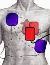
DSD is performed by attaching two sets of defibrillation pads rather than one and delivering two shocks as near simultaneously as possible, delivering electricity to the myocardial tissue in parallel pathways. The idea is that several factors affect the defibrillation threshold such as obesity, chronic lung disease, antiarrhythmic agents, decreased ejection fraction, body position/habitus, and presence of implanted internal defibrillator.

Hoch et al advocate for DSD in refractory VF. Hoch found that all five patients in the study converted to normal sinus rhythm after double sequence defibrillation at a total of 720 J. Other support for DSD come from the Cabanas paper, a retrospective case series which looked at 10 cases of refractory VF. In the paper, DSD successfully terminated 70% of refractory VF, attaining ROSC in 30% of those patients. Unfortunately, however, none of these patients survived to discharge. A contributing factor to explain the fact that there were no survivors to discharge was that DSD was performed too late. In the cases reviewed, 6.5 single shocks were given prior to DSD and in 6 of those cases, DSD was performed 35 minutes into resuscitation, which was probably too late.
           
Currently several systems around the world are using DSD for refractory VF.  Currently, we do not know the amount of joules to use for best survival. Nor do we know the correct number of pads or best pad vector. The risk/benefit profile seems very reasonable since all refractory VF leads to death. It is possible that we have finally figured out how to save these patients’ lives.

Anterior-Lateral/Anterior-Lateral

 Anterior-Lateral/Anterior-Posterior


References

  1. Chang, Mau-Song et al. Double and Triple Sequential Shocks Reduce Ventricular Defibrillation Threshold in Dogs With and Without Myocardial Infarction. Journal of the American College of Cardiology 1986; 8 (6): 1393-1405.
  2. Hoch, David H et al. Double Sequence External Shocks for Refractory Ventricular Fibrillation. JAC 1994; 23(5): 1141-1145.
  3. Zipes, Douglas P et al. Management of Patients with Ventricular Arrhythmias and the Prevention of Sudden Cardiac Death. American Heart Association, American College of Cardiology Foundation 2006.
  4. Pantridge, J. F et al. Electrical Requirements for Ventricular Defibrillation. British Medical Journal 1975; 2: 313-315.
  5. Geddes, L. A. et al. Electrical Dose of Ventricular Defibrillation of Large and Small Animals Using Precordial Electrodes. Journal of Clinical Investigation 1974; 53(1): 310-319.
  6. Adgey, A. A. J. Electrical energy requirements for ventricular defibrillation. British Heart Journal 1978; 40: 1197-1199.
  7. Cabaas, J. G. Double sequence external defibrillation in out-of-hospital refractor ventricular fibrillation: a report of ten cases. Prehospital Emergency Care 2015; 19(1): 126-130.
  8. Tacher, W. A. et al. Energy dosage for human trans-chest electrical ventricular defibrillation. New England Journal of Medicine 1974; 290: 214-215

Wednesday, April 8, 2015

The Tale of the Perfect Intubation

Disclaimer:  This is for the critically ill patient who is not in cardiac arrest.  Follow local protocols.  However, we believe this is the perfect technique for intubation.

1.      Place the patient on a High-Flow Nasal Cannula (HFNC) on at least 15 LPM.
2.      Put a non-rebreather on the patient over the nasal cannula at 15 LPM.
3.      Begin assessment for difficult airway and prepare suction.
4.      Put a PEEP valve on the BVM.
5.      Place the BVM attached to 100% O2 and PEEP valve over the patient’s face and remove the non-rebreather.
6.      Insert IV and begin IVF bolus running wide open, unless patient is in overt CHF.
7.      Give push-dose epinephrine at 10μg per minute if SBP <90 mmHg prior to intubation. Attempt to maintain SBP>90 mmHg at all times.  Alternative:  Start a “dirty” epinephrine drip (1 mg of code cart epi in 1 L of NS) run wide open. Typical flow rate of 18-20 gauge is 30cc/min = 30μg/min.
8.      If RR>4 and SpO2 >93%, do not bag!  Allow three minutes for denitrogenation/pre-oxygenation prior to intubation.
9.      If RR>4 and SpO2 <93%, do not bag!  Allow three minutes for denitrogenation/pre-oxygenation.  If SpO2 does not come up to 93% after three minutes, increase PEEP on PEEP valve, ensure proper positioning (see below), perform jaw thrust, and consider nasal airway (NPA).  Begin bagging patient at 6 breaths per minute (not more).  If pulse ox does not increase after three minutes of denitrogenation/pre-oxygenation followed by three minutes of BVM ventilation, perform rapid sequence intubation (RSI).
10.  If RR<4 and SpO2 >93%, ventilate at 6 breaths per minute and consider other causes, such as opioid overdose.  Consider “Rapid Sequence Airway” (supraglottic airway/iGel, decompress stomach, and gentle bagging).
11.  If RR<4 and SpO2 <93%, ventilate at 6 breaths per minutes until saturation at least 93%, then use BVM/PEEP valve without bagging for three minutes, then perform RSI.
12.  Ensure proper positioning.  This includes the head of bed at 20° elevation with ear aligned to the sternal notch.
13.  Give ketamine at 2 mg/kg.
14.  Give succinylcholine (if not contraindicated) at 1.5 mg/kg (or 2 mg/kg if SBP <90 mmHg).  Give rocuronium at 2 mg/kg, if available, in place of succinylcholine.
15.  Give fentanyl at 3 μg/kg for patients with head trauma.
16.  Perform laryngoscopy and intubation.
17.  Give fentanyl bolus at 2 μg/kg, followed by infusion at 1.5 ug/kg/hr, or repeat bolus if infusion not accessible.
18.  Repeat ketamine at 1 mg/kg PRN.
19.  Ventilate at 6 cc/kg (if ventilator available) and elevate the head of the bed/stretcher to 30°.

Wednesday, March 25, 2015

Status Epilepticus: A Ketamine-Deficient State?

Status epilepticus (SE) is a life threatening emergency associated with high morbidity and mortality rates. The International League Against Epilepsy (ILAE) defined SE more than twenty years ago as a single seizure that that lasts more than 30 minutes. The alternative definition is a series of epileptic seizures during which the patient’s baseline function is not regained between ictal events, within a 30 minute period. Recently, status epilepticus has been re-defined as: ≥5 minutes of continuous seizures OR ≥2 discrete seizures during which there’s an incomplete recovery of consciousness. Refractory status epilepticus (RSE) is defined as generalized or complex partial seizure activity that is refractory to conventional therapies for seizure disorder, such as benzodiazepines and barbiturates, within 30 minutes.  Super-refractory status epilepticus (SRSE) is further defined as SE that remains refractory to therapy with general anesthesia, for 24 hours, using medications such as propofol.  Incidence of refractory epilepsy is surprisingly high despite the development of many anti-epileptic agents and ranges from 20-40% in the literature.  Multi-faceted factors likely contribute to the development of RSE, including: the type of seizure, any underlying health conditions or neurological disorders, patient’s personal seizure history (frequency, duration, medication compliance, etc.), a patient’s genetics impacting drug metabolism (rate of absorption, metabolism, etc), or any prior use of illicit drugs altering brain chemistry (for example recreational MDMA, chronic alcohol or benzodiazepine use), amongst many others.  To prevent cortical disruption and damage, and to prevent morbidity and mortality, early control of SE is desirable.  Patients with development of RSE have high mortality rates (reported to approach 50%), increased hospital stays, poor functional outcomes, and inability to return to pre-admission baseline functional status.

Conventional therapies of acute seizure, SE, refractory and super refractory status epilepticus are heavily reliant on GABA agonist mediated therapies.  GABAa agonists control excitatory inhibition and spread of excitatory discharge.  Lorazepam and midazolam are listed to have Level A evidence for use as first line anti-seizure medications.  Second line therapies include sodium channel blockers such as phenytoin, fosphenytoin, levetiracetam, lamotrigine, and carbamazepine.  Also studied therapies include barbiturates, valproate, topiramate, and propofol.  Refractory states of SE are theorized to be attributable to alterations in receptors and disruption of molecular transport at the blood-brain barrier.  GABAa receptors are thought to be down-regulated with prolonged use of GABAa agonists. GABAa subunits are thought to undergo structural changes leading to impaired binding of anti-epileptic medications.  In addition, p-glycoprotein molecules are thought to be up-regulated as molecular transporters leading to increased efflux of medications from the brain, in particular phenytoin and phenobarbital.

With prolonged down regulation of GABAa receptors, an inhibitory function of excitation is lost. With decreased expression of GABAa receptors, increased expression and mobilization of non-competitive N-methyl-D-aspartate (NMDA) receptors to the cell surface of neurons occurs. Activation of NMDA receptors by glutamate will increase intracellular calcium, cause neuronal excitation, and potentiate refractory seizure physiology. In addition, prolonged treatment of RSE with traditional GABAa agonists may lead to development of refractory hypotension and other adverse cardiovascular effects. Hypotension in status epilepticus causes further insult to injury as seizures already have potential to cause anoxic damage as cerebral blood flow autoregulation is disrupted during seizures.

For all of these aforementioned reasons, conventional therapies can adversely impact treating SE. Ketamine has been postulated as a novel agent in treatment of RSE and perhaps has a role for early use in SE. Ketamine is a non-competitive NMDA glutamate receptor antagonist. Animal studies have demonstrated that ketamine is effective in control of refractory seizures and is neuroprotective (leading to decreased morbidity and mortality), when compared to control data. A recent systematic review of the literature by Zeiler (2015) summarized data of multiple human case reports and three prospective cohort studies (with an average of 7 patients per study) utilizing ketamine for RSE. Seizure resolution was established in 56% of 110 total adult patients and 63% of pediatrics patients. Most patients were with seizure resolution within 48 hours to 72 hours of start of ketamine. Treatment time ranged from 2 hours to 27 days in adults and 6 hours to 27 days in pediatrics. The literature is low powered and therefore statistically it is difficult to make generalizations to extrapolate its use and benefits for the general patient population. Dosing, duration, and outcomes with use of ketamine in RSE have been reported to vary, as well. Ketamine has sympathomimetic properties preventing hypotension and cardiac depression. It has been found to be especially useful in RSE when other anti-epileptic treatments are causal in cardiac depression or hypotension, eliminating need for pressor support with ketamine infusion. Treatment doses of continuous ketamine infusions range in the literature from 0.12mg/kg/hour to 10mg/kg/hour. At times, patients were initially bolused at doses ranging from 0.3mg/kg to 4.5mg/kg. Ketamine can be administered with low risk to most patients as there are few contraindications or adverse effects. Previous arguments for increased intracranial pressure with use of ketamine have been refuted in the literature, and few population groups exist where ketamine cannot be used safely for RSE. However, the prolonged use of ketamine and its effects have not been studied in the literature.

Prospective studies are required to help establish a role for NMDA antagonists in treatment of routine seizures. In addition, research should consider studying the use of ketamine earlier in treatment of seizure disorder. In one case report by Kramer (2012), ketamine was administered early on in treatment of SE after the patient began to develop worsening hypotension with escalating doses of midazolam and propofol. Ketamine infusion resulted in immediate reduction in prevalence, duration, and amplitude of seizures on EEG. The need for vasopressor support was weaned off and patient's seizures resolved within 12 hours of start of ketamine. Patient was discharged home with return to baseline level of function.

Now we potentially have another reason to use more ketamine!!!

References


F. A. Zeiler, “Early Use of the NMDA Receptor Antagonist Ketamine in Refractory and
Superrefractory Status Epilepticus,” Critical Care Research and Practice, vol. 2015, Article ID 831260, 5 pages, 2015.

Shorvon S. and Ferlisi M. “The treatment of super-refractory status epilepticus: a critical review of available therapies and a clinical treatment protocol.” Brain: A Journal of Neurology. 10. (2011):1-17.

Synoweic, et al. “Use of ketamine in the treatment of refractory status epilepticus.' Epilepsy Research. 105. (2013). 183-188.

Williams, et al. “Use of ketamine for control of refractory seizures during the intraoperative period.” Journal of Neurosurgical Anesthesiology. 26. (2014). 412.

Kramer AH. “Early Ketamine to Treat Refractory Status Epilepticus.” Neurocritical Care Society. 16. (2012): 299-305

Thursday, March 19, 2015

Let Apneic Oxygenation Reign!

We have talked about this topic briefly in the past but it is extremely important and deserves more dedicated attention.  Past mantra has dictated using a bag-valve mask (BVM) whenever a patient was thought to not be breathing adequately, or even not breathing at all.  Current evidence emphasizes the danger of the BVM and its inappropriate use.  The BVM can be summarized nicely: 1) It increases intrathoracic pressure thus decreasing preload and coronary artery perfusion, 2) opens the lower esophageal sphincter(even if you are good at ventilating) causing a high risk for vomiting from gastric insufflation, 3) causes over-distention of the alveoli resulting in oxygen shunting and decreased capillary PaO2. and 4) decreases cerebral blood flow. So what can we do to ensure oxygenation in those patients who just don’t require a BVM?  Apneic oxygenation!

The most common time the apneic oxygenation strategy will be employed is during the preoxygenation period and peri-intubation period of airway management.  The principles of apneic oxygenation may also be applied to patients who require supplemental oxygen but may not be able to be intubated at the time (i.e., predicted difficult airway or RSI meds are not available).  The goal is to maintain a SpO2 > 93% without using a BVM.  If the patient is unconscious during preoxygenation, this can be accomplished with a nasopharyngeal airway (NPA), nonrebreather (NRB) and high-flow nasal cannula (HFNC) set to at least 15 Lpm.  To review, the purpose of preoxygenation is to provide nitrogen washout.  As we know, nitrogen is the most common atmospheric gas and also predominates in your lungs.  If you remove the nitrogen by flooding the airway, including the dead space, with 100% O2 you can increase the functional reserve capacity and buy yourself time and reassurance during the apneic period of intubation. 

The best way to provide oxygenation during the apneic period of intubation is obviously to continue oxygenation.  Place an NPA (or two, yes two, NPAs) and passively oxygenate past the tongue through the glottis.  HFNC may actually provide bubbles or assist in visualization of the trachea; voila you have your view!  Through the use of HFNC during the apneic period of intubation the alveoli will continue to take up oxygen despite a lack of diaphragmatic movement or lung expansion.  Ideally the patient should remain in the upright 20 degree position ensuring that the airway remains patent with an NPA, OPA, jaw-thrust, head-tilt-chin-lift or, preferably, a combination to allow oxygen gas to pass down the nasopharynx into the deeper airway structures and then to the alveoli for passive diffusion.

Recent evidence has reviewed the effectiveness of the HFNC when used in the apneic period of intubation using RSI.  The study was conducted by an Australian helicopter emergency medical service (HEMS).  The HEMS service consisted of a physician and paramedic.  Intubation attempts were split evenly between the physician and paramedic in the pre-intervention arm of the study but favored the paramedics during the institution of apneic oxygenation.   They reviewed RSI intubations pre and post implementation of an apneic oxygenation protocol.  They had a significant decrease in desaturation during intubation in the group that received apneic oxygenation (22.6% to 16.5%).  They also noted a decrease in cardiac arrests (5.6% to 1.4%) and episodes of bradycardia (7% to 1.4%) related to desaturation during intubation after apneic oxygenation was implemented.  In conclusion, avoid the BVM whenever possible and always use a HFNC during intubation. 

So here is what I do EVERY TIME I am getting ready to intubate.  (Not in this order)
  1. Have a BVM ready with PEEP valve on at all times. 
  2. Have suction ready. 
  3. Quantitative ETCO2 ready to place on ETT.  (Remember, it needs to “zero” to the atmosphere first anyway.)
  4. Nasal cannula at least 15 LPM or as high as you can go. 
  5. Apply NRB at 100% over nasal cannula
  6. Bag only if RR<4 and SpO2 <93%.  If RR>4 and SpO2 <93% use oxygen for 3 MINUTES to see if saturation will come up.  If it does not come up… you can ventilate with BVM slowly (no more than 6 times per minute)


Bonus:  If systolic BP is less than 90 mmHg, add…
  1. IV fluid bolus as fast as possible
  2. Push-dose Epi 10 µg/min until SBP >90 mmHg


References
Weingart, SD. “Preoxygenation, Reoxygenation, and Delayed Sequence Intubation in the Emergency Department.” J Emerg Med 2010.
Weingart, SD, Levitan, RM. “Preoxygenation and Prevention of Desaturation During Emergency Airway Management.” Ann Emerg Med 2011.
Wimalesena Y, Burns B, Reid C, Ware S, Habiq K. “Apneic Oxygenation Was Associated With Decreased Desaturation Rates During Rapid Sequence Intubation by an Australian Helicopter Emergency Medicine Service.” Ann Emerg Med 2014. 

Wednesday, March 11, 2015

Let It Flow! Intraosseous Flow Rates by Insertion Site

Intraosseous (IO) access has become increasingly popular as a ‘safety net’ for failed IV access and has become a go-to procedure in pre-hospital cases of rapidly decompensating patients. Think of the cardiac arrest patient or the hemodynamically unstable trauma patient. The IO has proven a quick and reliable way to gain access to medullary venous plexuses in long bones, which drain into systemic venous circulation. We can basically think of the bone marrow as a vein that will not collapse on us that can be accessed very rapidly, with very little training. We have infused fluids, medications, and blood products successfully through the IO. And now that increased support has grown for the use of IOs prehospitally, the question has become which site is best: Tibia (which many people seem to be most comfortable with due to prominent landmarks and distance from resuscitative efforts), humerus, or sternum. There is literature supporting, and widespread consensus for, proximal tibia as the optimal insertion site in children, but this consensus does not exist in adults. Choice often depends on comfort level of the operator and convenience of the location, but we should also consider the difference in flow rates between sites.

There are few studies comparing IO placement sites but the ones that exist compare proximal to distal tibia, tibia to humerus, and one study which compared IO infusion rates between tibia, humerus and sternum in cadavers. The first study found that IO flow rates in the proximal tibia were significantly faster with and without use of a pressure bag than flow rates the in the distal tibia. The drawback of this study was its small sample size of only 22 patients. Pasley et al did a cadaver study published 1 year ago which utilized 16 cadavers to compare flow rates and found that the sternal site had the highest and most consistent flow rate compared to the humerus and tibia. In fact the average flow rate in the sternum according to this study was 1.6x higher than the humerus and 3.1x higher than the tibia. Additionally, this study showed that the tibia had the greatest number of insertion difficulties (In 3 out of the 16 cadavers, infusion was unsuccessful after insertion and alternate tibia had to be used.) Ong et al did a study in 2009 which had very different results. This study recruited 24 patients who presented to an ED in Singapore, all patients received a tibial IO, and those who needed a second access point were given a humeral IO (which 11 patients received). This study found no significant difference between the flow rates at the tibial and humeral site in contrast to Pasley’s study which did show a significant difference between humerus and tibia with the humeral site achieving a 1.8x greater volume on average than the tibia. Small sample size is an issue in all articles existing on this subject.

It seems that there have been no conclusive studies in human or cadaver studies on best IO insertion site, but, if we believe the most recent study by Pasley et al, the sternal and humeral IO sites, in that order, have better flow rates compared to tibial placement. This higher flow rate could make a difference when rapid fluid resuscitation is imperative and could lead to better survival of our patients. A new device called the FASTResponder was released by Pyng Medical in 2013 to make the sternal IO concept easier. This device is safe on ages 12 years and older and makes site identification easy. Another benefit of the device is, unlike the IO drill system, it requires no batteries, and, anecdotally, there is less pain on fluid delivery compared to other sites. One factor we are still unclear about is if the sternal IO could pose a problem if cervical immobilization is being used in trauma patients, with chest compressions, and for some airway procedures. Pyng Medical advertises on their website that it is “safe” to use in conjunction with cervical immobilization devices and CPR. However, the drill-based EZ-IO is approved for all ages, and many providers are already comfortable with it. According to Pasley’s study, the humeral placement is second best in terms of flow rates and had less insertional difficulties. He also notes that the humeral site had the greatest variability in volumes infused from subject to subject. There doesn’t seem to be enough evidence yet to draw firm conclusions; more studies are needed with a greater number of test subjects to increase reliability.  Furthermore, outcome measures, though often difficult to study, would be nice.

References

Carness J, Russell J, Rodrigo M, et al. Fluid Resuscitation Using the Intraosseous Route: Infusion with Lactated Ringer’s and Hetastarch. Military Medicine 2012; 2:222.

Ong M, Chan Y, Jen J, Ngo A. An observation prospective study comparing tibial and humeral intraosseous access using the EZ-IO. Amer Journal of Emergency Medicine 2009; 27, 8-15. 

Pasley J, Miller C, Dubose J, et al. Intraosseous Infusion Rates under High Pressure: A Cadaveric Comparison of Anatomic Sites. Distribution A: Approved for Public Release 2014: Case Number 88ABW-2014-1139.

Tan B, Chong S, Koh Z, Ong M. EZ-IO in the ED: an observational, prospective study comparing flow rates with proximal and distal tibia intraosseous access in adults. Amer Journal of EM 2012;30(8):1602-6.

Tuesday, February 10, 2015

Critical Illness Polyneuropathy- An Important Succinylcholine Contraindication

You are requested to your local short-term rehabilitation center for a 67 year old male with respiratory distress.  You arrive to find a patient in significant respiratory distress and altered mental status.  Per staff, the patient sustained an ischemic stroke ten days ago, which resulted in left-sided hemiparesis and some swallowing difficulties.  He has a past medical history of coronary artery disease and hypertension.  This morning, the patient developed acute respiratory distress and 911 was called.  You are concerned about pneumonia or pulmonary embolism in this bed-bound patient, and at this time it does not appear that he is protecting his airway.  Vitals are notable for BP 98/58, HR 128, sinus tachycardia on the monitor, respirations 40, and pulse oximetry 84% on RA.  You call medical control for delayed sequence intubation orders.  What regimen would you like to request?

After multiple clinical pearls on the topic summarizing the latest evidence, there should be little debate on the induction agent for this hypotensive patient.  Etomidate should be avoided in the hypotensive patient in extremis.  You request ketamine for induction sedation for this patient.

How many of you would choose succinylcholine for this patient?  Probably most of the readers would choose succinylcholine.  As you know, succinylcholine exerts its effects by depolarizing the neuromuscular junction by activating acetylcholine receptors.  The succinylcholine continues to activate the receptors, preventing repolarization, or a resetting, of the neuromuscular junction.  The effect continues until pseudocholinesterase, an enzyme in the body, metabolizes the succinylcholine.  The action of the depolarization does cause a potassium ion flux into the blood, typically no more than 1 mEq/L, even in instances of acute renal failure (e.g., dehydration, diabetic ketoacidosis).1

Most clinicians can rattle off the typical contraindications to succinylcholine administration, such as renal failure/hemodialysis, crush victims, burn victims, and prolonged immobilization/"found down."  Hopefully, if there is enough time to obtain a history, the question of, “Have you or anyone in your family had any problems with anesthesia in the past?” is being asked to ascertain the possibility of the very dangerous malignant hyperthermia.  If you’re really good, you may know that patients with myopathies, such as muscular dystrophy, may result in an acute rhabdomyolysis syndrome from the sudden muscle contractions of the depolarization process.  This may result in a sudden increase in serum potassium.  In fact, there is a black box warning on succinylcholine for this phenomenon, particularly in the pediatric population in which the myopathy may not yet be diagnosed in the patient.2

Much less known, though, is the critical illness polyneuropathy (CIP).  This clinical entity is seen primarily in ICU patients and patients with acute denervating injuries, such as a spinal cord injury or cerebrovascular accident.  In response to the sudden lack of nerve impulses coming from the upper motor neurons (i.e., the brain or spinal cord), the body starts to upregulate, or increase, the number of acetylcholine receptors at the neuromuscular junction in an attempt to make them more sensitive to any nerve signals coming their way.  While the body is unable to activate these neuromuscular junctions due to a functional blockade (e.g., severed spinal cord, ischemic area of brain), succinylcholine can still activate these junctions.  Since there are many more receptors, the activation of them will result in a greater flux of potassium out of the cells.  Potassium increases of 5-15 mEq/L have been seen in these instances, which can certainly cause cardiac arrest.  Because there is a delay in the production of additional receptors, the first 24 hours after an acute neurologic injury is typically safe for succinylcholine, so this should not change your practice with acute strokes.  The risk peaks 5 to 15 days after the denervating injury, and it is believed to last for 2-6 months afterwards.  However, some clinicians believe any patient with a history of denervating injury to be at risk for life-threatening hyperkalemia after succinylcholine.1,3–5

If you didn’t know this, you’re not alone.  After some clinicians in the UK had two hyperkalemic cardiac arrests in patients like this in their ICU after using succinylcholine, they surveyed other physicians who would be familiar with emergent intubations.  They found that 68.7% of survey respondents chose succinylcholine for intubation.6

To summarize, true contraindications to succinylcholine remain renal failure (particularly on hemodialysis), burns (cardiac arrests have occurred with as little as 8% body surface area involved), crush injuries, prolonged immobilization (e.g., found down at home and concern for rhabdomyolysis), myopathies, history of malignant hyperthermia, and, now, recent history of acute denervating injury, such as CVA or spinal cord injury.1

Case resolution:  You intubate the patient using ketamine and rocuronium, and you administer fentanyl and ketamine for post-intubation sedation.  The patient’s vital signs improve mildly.  At the emergency department, he is found to have a large saddle pulmonary embolus on CT angiography.  He goes to interventional radiology for thrombectomy (removal of the clot), as he cannot receive tissue plasminogen activator (tPA) due to the recent ischemic stroke.  His cardiodynamics improve significantly and he is extubated on hospital day #3.  He returns to rehab, albeit on a different regimen of anticoagulation.

References

1. Stollings JL, Diedrich DA, Oyen LJ, Brown DR. Rapid-sequence intubation: a review of the process and considerations when choosing medications. Ann. Pharmacother. 2014;48(1):62-76. doi:10.1177/1060028013510488.
2. Sandoz Inc. ANECTINE- succinylcholine chloride injection, solution (package insert). 2012. Available at: http://dailymed.nlm.nih.gov/dailymed/drugInfo.cfm?setid=579ff759-3099-45f5-befe-c4b79106c87e. Accessed September 21, 2014.
3. Biccard BM, Grant IS, Wright DJ, Nimmo SR, Hughes M. Suxamethonium and critical illness polyneuropathy. Anaesth. Intensive Care 1998;26(5):590-591.
4. Mallon WK, Keim SM, Shoenberger JM, Walls RM. Rocuronium vs. succinylcholine in the emergency department: a critical appraisal. J. Emerg. Med. 2009;37(2):183-8. doi:10.1016/j.jemermed.2008.07.021.
5. Booij LH. Is succinylcholine appropriate or obsolete in the intensive care unit? Crit. Care 2001;5(5):245-6.
6. Hughes M, Grant IS, Biccard B, Nimmo G. Suxamethonium and critical illness polyneuropathy. Anaesth. Intensive Care 1999;27(6):636-638.

Wednesday, February 4, 2015

Permissive Hypotension

You are dispatched to a scene where a 21 year old male has been shot in the chest by an unknown caliber handgun. On exam, you note a single GSW to the chest inferior to the left nipple. His VS are: BP 62/48, HR 138/min, RR 36 and labored. He is agitated and diaphoretic, but is AAOx4. You establish peripheral access and begin administering crystalloid fluid as a bolus. Your transport time to the trauma center is 20 minutes, due to road closures.
How much fluid should you administer en route to the trauma center?
            Captain Walter Cannon introduced the world to the concept of permissive hypotension in penetrating trauma back in 1918 during World War I. Cannon was an Army surgeon who witnessed the poor outcomes of patients who were “resuscitated” to “normal” blood pressures and developed the idea of the tenuous clot.  In 1994, Bickel and colleagues compared low volume resuscitation (300-340 ml) with standard ATLS volumes of 2400 ml.  In a randomized prospective trial, Bickel demonstrated a change in mortality of almost 7% in the low volume resuscitation group. This group also had less complications (ICU length of stay, development of acute respiratory distress syndrome, and abdominal compartment syndrome) compared with the standard resuscitation group.
            This practice of permissive hypotension in penetrating chest trauma is now widely accepted and practiced. The idea of the tenuous clot is real. Increased fluid volumes raise the blood pressure to levels higher than required, resulting in dilution of clotting factors and increased bleeding. The majority of these injuries are in non-compressible sites. Hence, patients end up bleeding more than they would have if we had never touched them in the first place.
            Therefore, consider resuscitating penetrating chest trauma patients to normal mental status.  This holds true for other trauma patients where bleeding is felt to be the cause of hypotension. The vast majority of people will retain normal mental status around 90 mmHg SBP. If the SBP is at least 90 mmHg, consider giving no fluids at all.

Boswell, K.  Menaker, J. Assessment and Treatment of the Trauma Patient in Shock, 2014-11-01Z, Volume 32, Issue 4, Pages 777-795, 

Saturday, January 31, 2015

Journal Club - February 2, 2015


  • What:  Weekly Journal Club
  • Where:  MONOC Education Building, 1415 Wyckoff Road, Ground Floor, Wall Township, NJ
  • When:  Monday, February 2, 2015 at 10:00 am

Live-tweeting of the journal club @CCareAnywhere.  #EMSJC

Friday, January 23, 2015

More Than Just Shock Value?

*Note - this discussion is only pertinent to modern biphasic defibrillators with self adhesive electrodes applied only anterior/posterior or anterior/lateral, with the compression provider wearing two pairs of gloves (double-gloving), with a maximum defibrillation energy of 360J.*
We have all heard the chant, “I’m clear, you’re clear, we’re all clear,” prior to a provider double, triple, sometimes quadruple checking him or herself before pushing that magic red button with the white lightning bolt - “shock”!  Recent literature has spurred quite the discussion on hands-on defibrillation (HOD) - CPR where compressions continue throughout the defibrillation - as it is widely known that interruptions in chest compressions lead to poor patient outcomes and are all too common, for example, during intubation, providing ventilations, AED analyzing, charging, and during defibrillation shocks.  This pearl is meant to provide a very brief explanation of what your risks might be, what protection devices you might use, anecdotal and published accounts on HOD, and suggestions for your clinical practice.
There are numerous factors in regards to energy and the effect it may have on the provider during HOD.  Energy is the product of voltage, current and time.  Neither factors, independently, are sufficient in inducing damaging effects.  For example, several thousand volts are experienced during static electricity, although the current is very low.  Current is determined by the resistance between the electrodes of the defibrillator, the electrode gel, the gel-skin contact, and the tissue resistance.  Glove integrity, skin moisture and the actual current pathway will determine the amount of escape current.  Biphasic defibrillators provide voltages up to approximately 2200 V over approximately 15-20 msec.  The maximum permissible leakage current, per the International Commission on Non-Ionizing Radiation Protection is 1mA; the threshold for perception is 2.5-4.0 mA; and pain is experienced at 6-10 mA.2  Sullivan and Chapman studied the voltage-current curves for gloves.  They note the international safety standard on 1mA and explain that at this level, it would take 1-3 seconds of current flow to induce VF in <5% of the population.  While defibrillation shocks are usually less than 20 msec, even if the pulse is timed appropriately in the rescuer’s cardiac cycle, as much as 500 mA would be required to induce VF.1 For reference, the current exposure from a home body fat monitoring scale is 500 uA.
In one of the most exciting studies, Lloyd et al measured current between “rescuers” and patients undergoing cardioversion at up to 360 J and found the highest current leak measured was 907 uA,4 with no “rescuers” experiencing a “shock.”  Neumann et al found HOD was safely performed on pigs by rescuers, HOD shortened pauses during CPR, and it more quickly restored coronary perfusion pressure.4  Kurz and Sawyer, in their letter to the editor of Resuscitation, advocate eliminating effects of no-flow time, perhaps by using HOD.7 Dr. Scott Weingart writes that in the 4 years that he and his colleagues have been performing HOD, there have been no rescuer complications, although occasional perceptions of tingling have been reported.  He himself reported arm soreness after 3 shocks, all at 360 J with the electrode pads notably in the anterior/anterior position.
In opposition, Lemkin et al derive an equation called the rescuer-received dose, to try to better qualify defibrillation risk.  Noting that energy values greater than 1 J reportedly can cause VF, they deem HOD unsafe as values above 1 J were calculated in their cadaver study, though effects of gloves were not accounted for.  Two studies from the UK found that medical examination gloves do not provide rescuer safety and even demonstrate further glove breakdown of the gloves worn by rescuers who perform compressions.  According to Sullivan and Chapman, HOD with medical examination gloves will produce no sensation at all unless the gloves completely break down.1  
Although there are no reported fatalities or serious consequences to rescuers performing HOD under ideal conditions - using a biphasic defibrillator with electrodes placed appropriately, with rescuers double gloved - we should take note that any change to a safety protocol should not be undertaken without ensuring rescuers' safety.  I have personally performed HOD, as have my colleagues in the emergency department.  While none of us have experienced any detrimental consequences or even the reported tingling, considering the literature, perhaps we should currently hold off on changing our protocols to mandate hands-on defibrillation.  Protocols that need to be changed or followed are as follows:

  • High quality CPR remains of utmost importance.  Set a metronome at 100 beats per minute and compress the chest to 1.8” (or as close to it as possible) every time.
  • Have no interruptions in chest compressions - not for intubation, not for starting an IV, not for inserting a central line, not for transporting, and not for charging the defibrillator!
                  
The use of HOD needs to reflect your clinical decision made in the best interest of you, your co-rescuers, and your patient.  If you chose to do so, please double-glove, please place the electrodes anterior/posterior, and communicate your practice to your colleagues.    

References

1.  Sullivan JL, Chapman FW. Will medical examination gloves protect rescuers from defibrillation voltages during hands-on defibrillation? Resuscitation. 2012 Dec;83(12):1467-72. doi: 10.1016/j.resuscitation 2012.07.031. Epub 2012 Aug 25. PubMed PMID: 22925991

2.  Petley GW, Cotton AM, Deakin CD. Hands-on defibrillation: theoretical and practical aspects of patient and rescuer safety. Resuscitation. 2012 May;83(5):551-6. doi: 10.1016/j.resuscitation.2011.11.005. Epub 2011 Nov 15. Review. PubMed PMID: 22094984.

3.  Sullivan JL. Letter by Sullivan regarding article, "Hands-on defibrillation: an analysis of electrical current flow through rescuers in direct contact with patients during biphasic external defibrillation". Circulation. 2008 Dec 2;118(23):e712; author reply e713. doi: 10.1161/CIRCULATION AHA.108.803718. PubMed
PMID: 19047587.

4.  Lloyd MS, Heeke B, Walter PF, Langberg JJ. Hands-on defibrillation: an analysis of electrical current flow through rescuers in direct contact with patients during biphasic external defibrillation. Circulation. 2008 May 13;117(19):2510-4. doi: 10.1161/CIRCULATION AHA.107.763011. Epub 2008 May 5. PubMed PMID: 18458166.

5.  A note of caution on the performance of hands-on biphasic defibrillation. Weingart SD. Resuscitation. 2013 Mar;84(3):e53. doi: 10.1016/j.resuscitation.2012.12.014. Epub 2012 Dec 22. PMID: 23266533

6. Lemkin DL, Witting MD, Allison MG, Farzad A, Bond MC, Lemkin MA. Electrical exposure risk associated with hands-on defibrillation. Resuscitation. 2014 Oct;85(10):1330-6. doi: 10.1016/j.resuscitation.2014.06.023. Epub 2014 Jun 30. PubMed PMID: 24992873.

7.  Petley GW, Deakin CD. Do clinical examination gloves provide adequate electrical insulation for safe hands-on defibrillation? II: Material integrity following exposure to defibrillation waveforms. Resuscitation. 2013 Jul;84(7):900-3. doi: 10.1016/j.resuscitation.2013.03.012. Epub 2013 Mar 16. PubMed PMID: 23507465.

8.  Deakin CD, Lee-Shrewsbury V, Hogg K, Petley GW. Do clinical examination gloves provide adequate electrical insulation for safe hands-on defibrillation? I: Resistive properties of nitrile gloves. Resuscitation. 2013 Jul;84(7):895-9. doi: 10.1016/j.resuscitation.2013.03.011. Epub 2013 Mar 16. PubMed PMID: 23507464.

Wednesday, January 7, 2015

Did you get the orthostatics yet?

A 70 y/o male presents from a nursing facility with symptoms of weakness after 2 days of diarrhea.  He states it has been watery and occurring 4-5 times per day.  His heart rate is 80 bpm and regular, BP 130/70, respirations of 16, skin warm and dry. He appears well but shows a little general weakness overall.  He knows he takes medications for his blood pressure; however, he is not sure of the name.

Q: Would you get orthostatic vital signs on this patient to assess for volume loss?

Orthostatic vital signs have been used to assess for volume loss by measuring the bodys response to positional change.  Upon standing from a supine position, vasoconstriction and changes in heart rate help to maintain perfusion.  It is thought that when a person is hypovolemic this system fails and blood pools in the lower extremities causing a drop in blood pressure and/or an increase in heart rate.   Symptoms of orthostatic hypotension are lightheadedness, dizziness, blurred vision, weakness, fatigue, cognitive impairment, nausea, palpitations, tremulousness, headache, and syncope.  Orthostatic vital signs are considered positive when there is a drop in systolic blood pressure of ≥ 20 mmHg, drop in diastolic blood pressure of ≥ 10 mmHg, or heart rate increase of ≥ 30 beats per minute within 3 minutes of standing from a supine position.1

The utility of orthostatic vital signs came into question over 20 years ago.  A study in 1990 looked at orthostatic vitals in 132 self-proclaimed euvolemic patients aged 18-80 years old (mean 34.1 +/- 13.6 years).  Of these patients 43% tested positive.  The study concluded that normal patients may present with orthostatic vitals given the current criteria.2

In 1997, a study examined orthostatics in 911 non-acutely ill patients aged greater than 60 from 45 different nursing homes.  To be included in the study, patients had to be able to stand for at least one minute.  The study found that over 50% of patients had orthostatic changes at baseline and it was most prevalent in the morning when patients first rise.3

Besides the elderly, orthostatic vitals were examined in adolescents as well.  307 healthy high school students aged 15-17 were checked for orthostatic vitals.  The study found pulse changes within the population to be 61% sensitive and 56% specific.  They also found orthostatic blood pressure changes to be within the adult range for 98% of adolescents, and a third of participants experienced orthostatic symptoms.  The study concluded the orthostatic heart rate criterion to be likely inappropriate for adolescents.4  Another study examining blood pressure changes in 23 healthy adolescents concluded transient orthostatic hypotension is common in their population.5

In addition to examining orthostatics in the non-acutely ill and adolescents, they were also studied in patients with known blood volume loss.  A study in 1992 examined 100 blood donors aged 19-83 years old and 100 senior center volunteers aged 55-94. The blood donors all gave 450 mL of blood.  Orthostatics had no clinical difference between ages.  Furthermore, a pulse rise >20 bpm or a diastolic BP drop > 10 mmHg had a specificity of 17%, sensitivity of 98%.  Systolic changes yielded no better.6 A similar study from 1994 looked at orthostatics in blood donation of 450 mL between two age groups, patients <65 and patients 65 or older.  These were healthy volunteers at baseline prior to blood donation.   A pulse change >20 bpm was found to have a sensitivity of 43% and a specificity of 94% in patients less than 65 years old.  In the age 65 and older group, pulse change was found to have a sensitivity of 25% and a sensitivity of 100%.  When they looked at blood pressure, they found it was worse than the flip of a coin.7

Besides blood volume loss, fluid volume loss and orthostatics were also studied. A study of 23 pregnant women with hyperemesis gravidarum studied the sensitivity of orthostatics in pre and post rehydration of 6 liters of lactated Ringers solution.  The study found that orthostatic changes lack sufficient sensitivity to be effectively used as quantitative screening tests for dehydration.8

In summary, the review above shows that using orthostatic vital signs alone to determine volume loss is highly unreliable.  Many patients can test positive for orthostatic signs even when asymptomatic.  We would never want to utilize a test that is so sensitive yet essentially with minimal specificity.  This would then cause the healthcare provider to act on all of the “positive” results by assuming the patient is hypovolemic. To make matters worse, the proportion of patients on beta blockers causing a blunting of the testing would make this even more unreliable than it already is.  When patients were known to have volume loss, orthostatic vitals still lacked a sufficient sensitivity to be deemed an effective test.  Looking for orthostatic clinical signs, not the numbers, is a far more reliable means to assess volume loss.  If the patient stands up and feels either lightheaded or passes out, this is sufficient enough to determine significant hypovolemia.  

References
1.         Naccarato M, Leviner S, Proehl J, et al. Emergency Nursing Resource: orthostatic vital signs. Journal of emergency nursing: JEN : official publication of the Emergency Department Nurses Association. Sep 2012;38(5):447-453.
2.         Koziol-McLain J, Lowenstein SR, Fuller B. Orthostatic vital signs in emergency department patients. Annals of emergency medicine. Jun 1991;20(6):606-610.
3.         Ooi WL, Barrett S, Hossain M, Kelley-Gagnon M, Lipsitz LA. Patterns of orthostatic blood pressure change and their clinical correlates in a frail, elderly population. Jama. Apr 23-30 1997;277(16):1299-1304.
4.         Skinner JE, Driscoll SW, Porter CB, et al. Orthostatic heart rate and blood pressure in adolescents: reference ranges. Journal of child neurology. Oct 2010;25(10):1210-1215.
5.         Stewart JM. Transient orthostatic hypotension is common in adolescents. The Journal of pediatrics. Apr 2002;140(4):418-424.
6.         Baraff LJ, Schriger DL. Orthostatic vital signs: variation with age, specificity, and sensitivity in detecting a 450-mL blood loss. The American journal of emergency medicine. Mar 1992;10(2):99-103.
7.         Witting MD, Wears RL, Li S. Defining the positive tilt test: a study of healthy adults with moderate acute blood loss. Annals of emergency medicine. Jun 1994;23(6):1320-1323.
8.         Johnson DR, Douglas D, Hauswald M, Tandberg D. Dehydration and orthostatic vital signs in women with hyperemesis gravidarum. Academic emergency medicine : official journal of the Society for Academic Emergency Medicine. Aug 1995;2(8):692-697.